1. What is the lumbar attachment of the diaphragm composed of?
a. Two aponeurotic arches: medial and lateral
b. Two arcuate ligaments: medial (is situated superior to the quadratus lumborum) and lateral (is situated superior to the psoas major)
c. Two crura or pillars
d. One wide, compact aponeurotic attachment
e. Only the two aponeurotic arches: medial and lateral
2. Name the features of the diaphragmatic central tendon:
a. Its center is formed by four strong diagonal bands
b. It has a trifoliate shape
c. It has a bifoliate shape
d. The hiatus for the passage of the inferior vena cava is located in this area
e. The hiatus for the passage of the aorta is located in this area
3. Name the diaphragmatic apertures:
a. Thoracic duct opening
b. Aortic opening
c. Splanchnic nerves opening
d. Inferior vena cava opening
e. Oesophagus opening
4. When the diaphragm contracts, does it affect the aorta?
a. Yes, because the aorta passes through the aortic hiatus when entering the abdomen so, the diaphragmatic muscle will constrict the hiatus
b. Yes, because the aorta passes through the aortic hiatus which is formed by the diaphragm on one side and the vertebral column on other side, the contractions of the muscle will push the aorta towards the vertebral column and minimize the diameter of the aorta
c. No, because the aorta passes through the aortic hiatus, which is a tendinous one, not muscular, and it’s located posterior to thexifoid process of the sternum
d. No, because the aorta has nothing to do with the diaphragm
e. No, because the aortic hiatus is located between the median arcuate ligament anterior, vertebral column posterior and diaphragmatic crura lateral, basically posterior the muscular part of the diaphragm, this way the contractions of the muscle do not affect the aorta
5. What passes through the aortic hiatus of the diaphragm?
a. Aorta
b. Phrenic nerves
c. Thoracic duct
d. Lymphatic trunks
e. Sometimes the azygos vein
6. What passes through the oesophageal hiatus of the diaphragm?
a. Phrenic nerves
b. Thoracic duct
c. Oesophagus
d. Vagal trunks
e. Gastric nerves
7. The oesophageal diaphragmatic hiatus has the following features:
a. Is located at the level of the twelve thoracic vertebra
b. Is located anterior, superior and lateral to the aortic hiatus
c. There is a distinct continuity between the oesophageal wall and the muscular fibres that form the hiatus
d. There is a loose connecting tissue between the inferior part of the oesophagus and the hiatus that permits movement of the organ when swallowing and ventilating
e. The only organ that passes through the oesophageal diaphragmatic hiatus is the oesophagus
8. The arterial supply of the diaphragm is insured by the:
a. Phrenico-oesophageal arteries
b. Superior and inferior phrenic arteries
c. Pericardiacophrenic and musculophrenic arteries
d. Phrenico-gastric arteries
e. Last five intercostal and subcostal arteries
9. What is the most common origin of the inferior phrenic arteries?
a. Right and left gastric arteries
b. Coeliac trunk
c. Oesophageal arteries
d. Aorta
e. Renal arteries
10. The venous drainage of the superior surface of the diaphragm is ensured by:
a. Superior right phrenic vein
b. Superior left phrenic vein
c. Tributaries of the musculophrenic veins
d. Tributaries of the pericardiacophrenic veins
e. Tributaries of the gastric veins
11. The innervation of the diaphragm is done by the:
a. Phrenic nerves
b. Vagus nerves
c. Lower six or seven intercostal nerves
d. Recurrent laryngeal nerves
e. Splanchnic nerve
12. What are the attachments of the external oblique muscle?
a. Origin on the external surfaces of 5th– 12th ribs
b. Insertion from superior to inferior-lateral: linea alba, pubic tubercle and anterior half of iliac
crest
c. Origin on the thoracolumbar fascia, anterior two thirds of iliac crest
d. Insertion on the external surfaces of 10th– 12th ribs
e. Insertion on the xiphoid process and 5th– 7th costal cartilages
13. What are the attachments of the internal oblique muscle?
a. Origin on the thoracolumbar fascia, anterior two thirds of iliac crest, iliopectineal arch (lateral two- thirds of the inguinal ligament)
b. Insertion on the inferior borders of 10th–12th ribs, linea alba
c. Origin on the inner surfaces of 7th– 12th ribs
d. Insertion on the xiphoid process and 7th– 12th costal cartilages
e. Origin on the external surfaces of 7th– 12th ribs
14. The anterior wall of the inguinal canal is formed by the:
a. Linea alba
b. Rectus abdominis muscle
c. Transversus abdominis muscle
d. Internal oblique
e. Aponeurosis of external oblique
15. The posterior wall of the inguinal canal it’s form by the:
a. The conjoint tendon
b. The transversalis fascia
c. Rectus abdominis muscle
d. External oblique
e. Peritoneum
16. What passes through the inguinal canal in man:
a. Spermatic cord
b. Round ligament
c. Ilioinguinal nerve
d. Iliohypogastric nerve
e. Femoral artery
17. What passes through the inguinal canal in women:
a. Inferior epigastric vessels
b. Ovarian arteries
c. Ilioinguinal nerve
d. Femoral vein
e. Round ligament
18. What is the linea alba?
a. The aponeurosis of external oblique
b. A tendinous strap that is located between the xiphoid process and pubic symphysis
c. A junction of fibers from the aponeurosis of external oblique, internal oblique and transversus abdominis muscle
d. A junction of fibers from the aponeurosis of external oblique, internal oblique and rectus abdominis muscle
e. A junction of fibers from the aponeurosis of external oblique, internal oblique, transversus abdominis muscle and rectus abdominis muscle
19. A patient has a very strong pain just inferior to the xifoid process, that is the name of that area?
a. Epigastrium
b. Hypochondrium
c. Umbilical
d. Lumbar
e. Hypogastrium
20. Which are the landmarks of the inferior border of the abdominal wall?
a. Anterior superior iliac spine
b. Posterior superior iliac spine
c. Greater sciatic notch
d. The inguinal ligament
e. The pubic tubercle and the pubic crest
21. What does linea semilunaris mean?
a. The curved area inferior to the costal ribs
b. Curved fold superior to the pubic symphysis
c. Shallow, curved groove lateral to lateral margin of rectus sheath
d. Curved aponeurosis inferior to the umbilicus
e. Curved aponeurosis superior to the umbilicus
22. What is the posterior rectus sheath made of?
a. Posterior lamina of internal oblique
b. Anterior lamina of internal oblique
c. Posterior laminae of external oblique
d. Posterior laminae of transversus abdominis
e. Anterior laminae of transversus abdominis
23. The inferior epigastric artery it’s a branch of the:
a. Femoral artery
b. Internal iliac artery
c. External iliac artery
d. Abdominal aorta
e. Common iliac artery
24. Arterial supply of the rectus abdominis muscle is done by the:
a. Lumbar arteries
b. Internal thoracic
c. Superior epigastric artery
d. Inferior epigastric artery
e. Umbilical artery
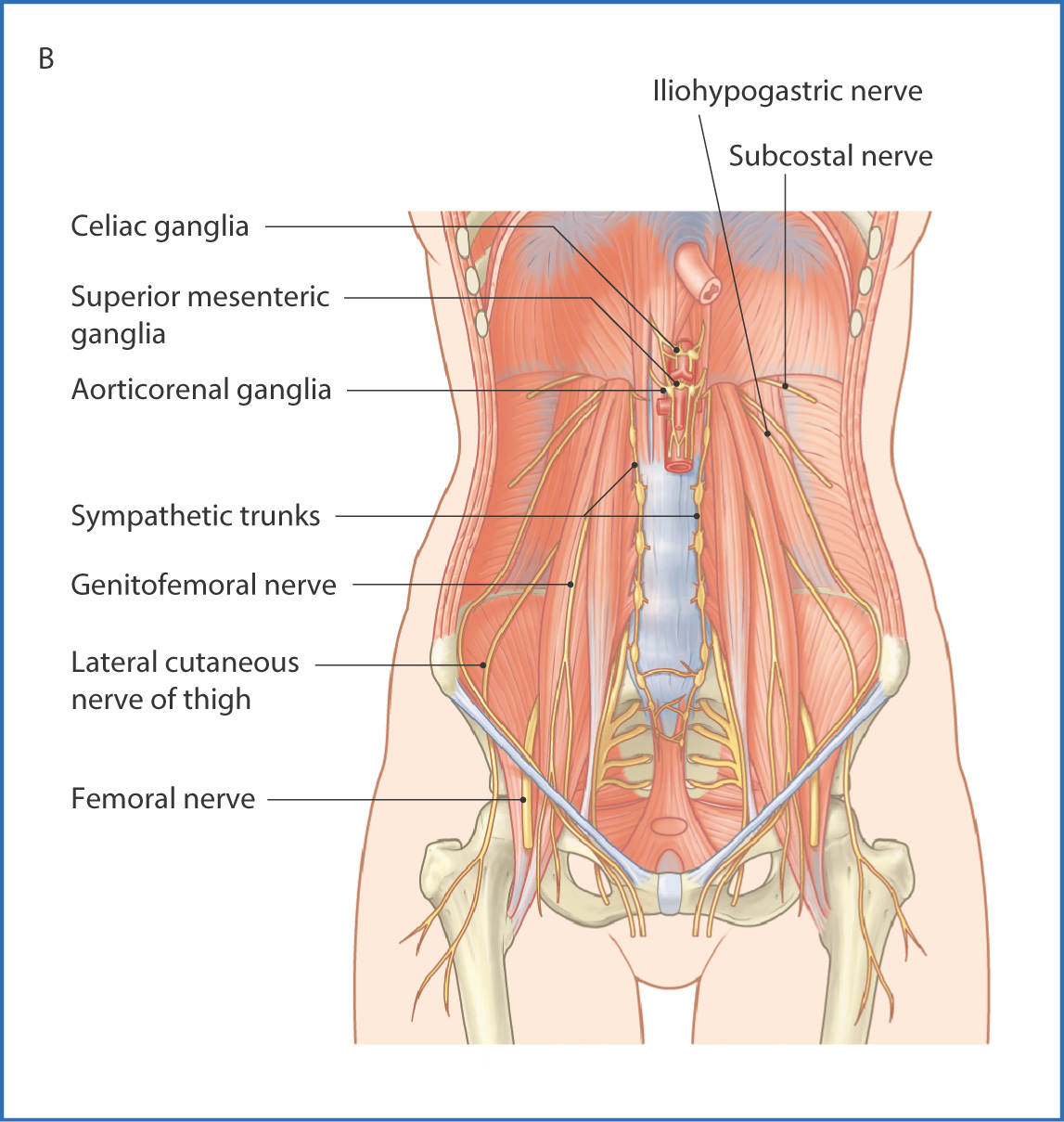
25. What nerves pass anteriorto the quadratus lumborum muscle?
a. Subcostal
b. Lumbosacral
c. Iliohypogastric
d. Ilioinguinal
e. Genitofemoral
26. Patient has a tumor located at the level of the right flank what muscles may be involved?
a. Rectus abdominis muscle
b. Transversus abdominis muscle
c. Internal oblique
d. External oblique
e. Pyriformis
Answers
1. a, c
2. a, b, d
3. b, c, d, e
4. e
5. a, c, d, e
6. c, d, e
7. b, d
8. b, c, e
9. b, d
10. c, d
11. a, c
12. a, b
13. a, b
14. d, e
15. a, b
16. a, c
17. c, e
18. b, c
19. a
20. a, d, e
21. c
22. a, d, e
23. c
24. c, d
25. a, c, d
26. b, c, d
